Electroretinography (ERG)
¿Por qué es importante la electrorretinografía?
¿Qué es la electrorretinografía (ERG)?
Electroretinography is an electrophysiological test of the retina, the layer of the eye which detects light. The electroretinogram (ERG) is to the retina what the electrocardiogram (ECG) is to the heart. Just as an ECG is crucial to diagnosing illness and monitoring the heart’s function, ERG plays a critical role in the care of the eye, and is instrumental in the early detection of retinal dysfunction.
¿Por qué el ERG es importante?
La electrorretinografía ofrece una gran cantidad de información vital y objetiva sobre la función y el estado de la retina. La ERG desempeña un papel fundamental en el diagnóstico de enfermedades oculares hereditarias y adquiridas, además de impulsar la investigación de los factores que afectan a la salud de la retina o de las vías visuales.
¿Qué es un electrorretinograma?
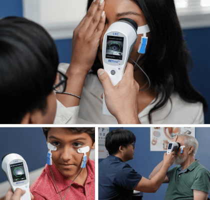
The electroretinogram (ERG) is an examination that evaluates the function of the eye’s retinal cells. Patients’ eyes are stimulated with light and the resulting electrical activity from their retinal cells is measured by skin or corneal electrodes. Dependent on the type, intensity and colour of the light stimulus information on different areas and types of cells can be obtained.
3 tipos de ERG
Existen tres tipos principales de ERG: ERG de campo completo (ffERG), ERG patrón (PERG) y ERG multifocal (mfERG). Cada uno de ellos tiene diferentes aplicaciones y beneficios. La determinación del estado funcional de la retina con diferentes tipos de ERG permite a médicos e investigadores evaluar a los pacientes para detectar afecciones relacionadas con la retina, monitorizar de forma fiable la función de la retina con el tiempo y evaluar la eficacia de los tratamientos de la retina. Los tres tipos principals de ERG se utilizan en la atención a personas, en clínicas veterinarias y en la investigación médica.

ERG de campo completo
(ffERG)
En el ERG de campo completo (ffERG), los electrodos registran la respuesta eléctrica sumada de las células de la retina. La respuesta eléctrica sumada es una combinación de los resultados de varias células y regiones diferentes de la retina, lo que significa que el ERG de campo completo (ffERG) muestra el funcionamiento de la retina en su conjunto. El dispositivo RETeval® manual y portátil es capaz de realizar ffERG.

Patrón ERG
(PERG)
En el ERG patrón (PERG), el estímulo de luz es un patrón de tablero de ajedrez que evoca respuestas eléctricas de las células ganglionares que forman la retina interna. El PERG se suele utilizar como prueba para enfermedades de las células ganglionares, así como muchas enfermedades neurodegenerativas.[8] El UTAS device™ offers PERG testing with custom protocols and bilateral testing capabilities.

ERG multifocal
(mfERG)
El ERG multifocal (mfERG) se lleva a cabo siguiendo un protocolo similar al del PERG pero el estímulo de luz forma un variedad de hexágonos que parpadean entre blanco y negro en una secuencia pseudoaleatoria. El mfERG es ideal para medir la disfunción de la zona central de la retina y no es sensible a anomalías del axón ganglionar[9] El dispositivo UTAS offers multifocal testing with different options for stimulus type and reporting of results.
Detectar estrés funcional
Anticipar daño estructural.
¿En qué se diferencia la electrorretinografía de una imagen estructural?
La electrorretinografía evalúa objetivamente las anomalías de la retina, mientras que las imágenes estructurales muestran la anatomía del tejido de la retina. Si bien tanto las evaluaciones funcionales como las estructurales tienen sus beneficios, los cambios funcionales generalmente aparecen mucho antes que los cambios estructurales. Detectar rápidamente las anomalías de la retina es fundamental para minimizar el daño y maximizar la retención de la visión, por lo que las evaluaciones funcionales como la ERG son herramientas importantes en el conjunto de diagnósticos oftalmológicos.
ERG portátil como herramienta de diagnóstico práctica y eficaz
Recent studies comparing ERG to structural imaging techniques show that ERG is an effective diagnostic tool. In studies comparing ERG and structural imaging’s abilities to evaluate sight-threatening diabetic retinopathy, the handheld and portable RETeval device’s ERG results outperformed the traditionally-used imaging technique in predicting which patients would later need medical intervention.[13],[14] En una evaluación de la capacidad de RETeval’s ability to evaluate diabetic retinopathy, the advantages of RETeval incluyeron la detección temprana de la disfunción retiniana, menores costos de inversión y menos conocimientos de lectura requeridos que la técnica de diagnóstico por imágenes utilizada tradicionalmente.[15] En una evaluación de la capacidad de RETeval’s ability to evaluate central retinal vein occlusion found that RETeval son menos invasivos, más fáciles de realizar y brindan predicciones más confiables que la técnica de imágenes que se usa tradicionalmente.[16]
Ventajas de las pruebas ERG
La facilidad y sensibilidad de los ERG realizados con el RETeval portátil es muy preferible a las técnicas de imagen en determinadas situaciones. Los pacientes que tienen opacidades mediáticas, mala cooperación o pupilas pequeñas pueden ser evaluados mejor para detectar anomalías en la retina con el RETeval[14].
Pruebas ERG no invasivas
En un estudio independiente en el que se compararon los ERG de RETeval con las fotografías digitales convencionales de la retina, los pacientes eligieron el RETeval como el método preferido sobre la fotografía.[14] ] Si bien muchos niños necesitan ser sedados para someterse a una ERG convencional, los niños de tan solo seis meses de edad han podido mantener la calma durante las ERG realizadas con RETeval.[18] La naturaleza no invasiva del ERG y la facilidad del RETeval portátil y no midriático hacen que el dispositivo ERG sea una valiosa herramienta de detección para muchas patologías oftálmicas.
RETeval, pruebas ERG simplificadas
Aplicaciones de la electrorretinografía
La electrorretinografía presenta una amplia variedad de aplicaciones en humanos y animales, tanto para uso clínico como de investigación. Nuestra tecnología ofrece soluciones de ERG fiables, reproducibles y efectivas en todas estas categorías.


More about the RETeval ERG/VEP device
Popular Topics: Make a Difference in Diabetic Retinopathy Care | Glaucoma Evaluation with RETeval PhNR Test | RETeval Device Reference Data | RETeval in Optometry
Case Studies: Photopic Negative Response as a Reliable Method for Glaucoma Follow-up in Children | A Tale of Two Patients | Vision Complaints Reflected on ERG | Predictive Value of Combining Diagnostic Technologies| ERG Provides Clarity When Fields and OCT Are Inconclusive| ERG Raises Red Flag, Changing Management Trajectory | ERG Provides Confidence to Monitor or Treat | ERG to Determine Ischemic Status | ERG to Replace FA for CRVO Treatment Decision | Using ERG to Monitor Glaucoma | Routine ERG Use Supports Complex Patient Management | ERG Alters Follow-up Schedule and Education for Patient with Diabetes | Using ERG for Management of Birdshot Chorioretinopathy | Using ERG to Monitor Glaucoma | Comprehensive Pediatric Assessment Using ERG in Challenging Cases | ERG Above and Beyond Retinal Imaging | ERG’s Role in Diabetic Retinopathy Progression Monitoring | Evaluación de riesgos basada en ERG en CRVO | ERG Supports Diagnostic Accuracy in a Pediatric Patient
Ebooks: Core Cases from the Clinical Compendium | Modern Fundamentals of Diabetic Retinopathy Management in Optometry | Elevating Patient Care with ERG
Articles: Electroretinography Added to AAO’s Diabetic Retinopathy Preferred Practice Pattern Guidelines | ¿Es cómodo el dispositivo RETeval para los pacientes? |The Use of RETeval ERG/VEP en oftalmología pediátrica | How RETeval ERG Has Enhanced My Practice | The Use of RETeval ERG/VEP in Pediatric Ophthalmology | The Ultimate Guide to Diabetic Retinopathy in Primary Eyecare | What Type of Functional Testing Do You Prefer for Patients with Diabetes? | Major Milestone: RETeval Referenced in over 200 Publications | Collaboration to Elevate the Standard of Care for DR | Is ERG Needed if You Have Access to a Good Structural Imaging Device? | UN ENFOQUE SENCILLO PARA EL MANEJO Y APOYO DE LOS PACIENTES CON DIABETES | Simplify Grading and Risk Assessment in Diabetic Retinopathy | Simplify Daily Decision-Making with Modern ERG | Objective Functional Testing Needs in Diabetes and Glaucoma | Why Modern ERG is Re-Defining Diabetes Management | Diabetic Retinopathy Management Protocols for Optometry
Videos: RETeval: Function to Rely On | RETeval Handheld ERG: Features & Benefits | RETeval: Enhancing Collaborative Care | Handheld ERG for Primary Eyecare | Advice for Optometric Colleagues about Handheld ERG | Making a Difference in Diabetic Retinopathy Care | Changing the Way We Think About Electrodiagnostics | ERG Testing Made Simple | VEP Testing Made Simple | ERG Waveform | Introduction to Visual Electrophysiology | A Superior DR Progression Risk Assessment with the RETeval manual y portátil, | Improve Glaucoma Management with the RETeval Handheld ERG Device | Reshaping the Retinal Diagnostic Landscape | New Solutions for Infants with ROP | Using the RETeval in Myopia Research
Webinars: ABCs of ERG | ERG in Action | Blueprint for Functional Assessments | Predicting Vision Loss in a Busy Retina Practice | Objective, Functional Testing for Glaucoma? | Best Management Practices for Diabetic Retinopathy | How the RETeval Device Became a Daily Instrument in my Diagnostic Toolkit
¡Hablemos!
Solicite una demostración o más información sobre nuestros dispositivos.