Caso de Estudio
ERG Demonstrates Stable Function Despite Severe Structural Damage
by Josué Roberto Lozano, MD, Centro Médico Hidalgo, Monterrey, N.L., Mexico

Desafío
Structure & Function Mismatch

DIAGNÓSTICO
Glaucoma

Testing Protocol
PhNR (Respuesta Fotópica Negativa)
Patient History
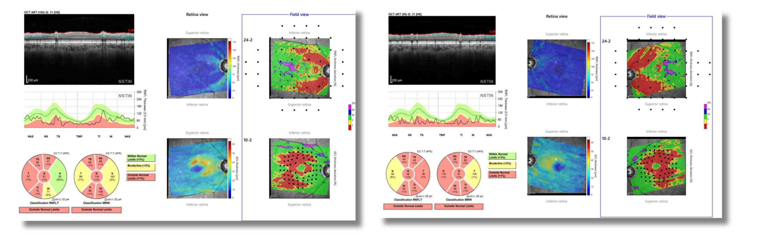
A 72-year-old male patient had been diagnosed with primary open-angle glaucoma for 4 years. He is pseudophakic in both eyes and has bilateral extended depth-of-focus intraocular lenses. The patient had previously been referred to a sleep clinic and diagnosed with obstructive sleep apnea. During routine follow-up, fundus photography (Figure 1) revealed marked structural damage in both optic nerves, including extensive cupping nearing 90%. The OCT (Figure 2) revealed prominent beta-zone atrophy, and classic bayonet vessel signs – despite maintaining good visual acuity (20/20 in the right eye and 20/25 in the left eye). His current treatment consisted of timolol, brimonidine, Ginkgo biloba 120 mg daily, and continuous CPAP therapy. However, obtaining a visual field was not available in this particular case.
Figure 1:
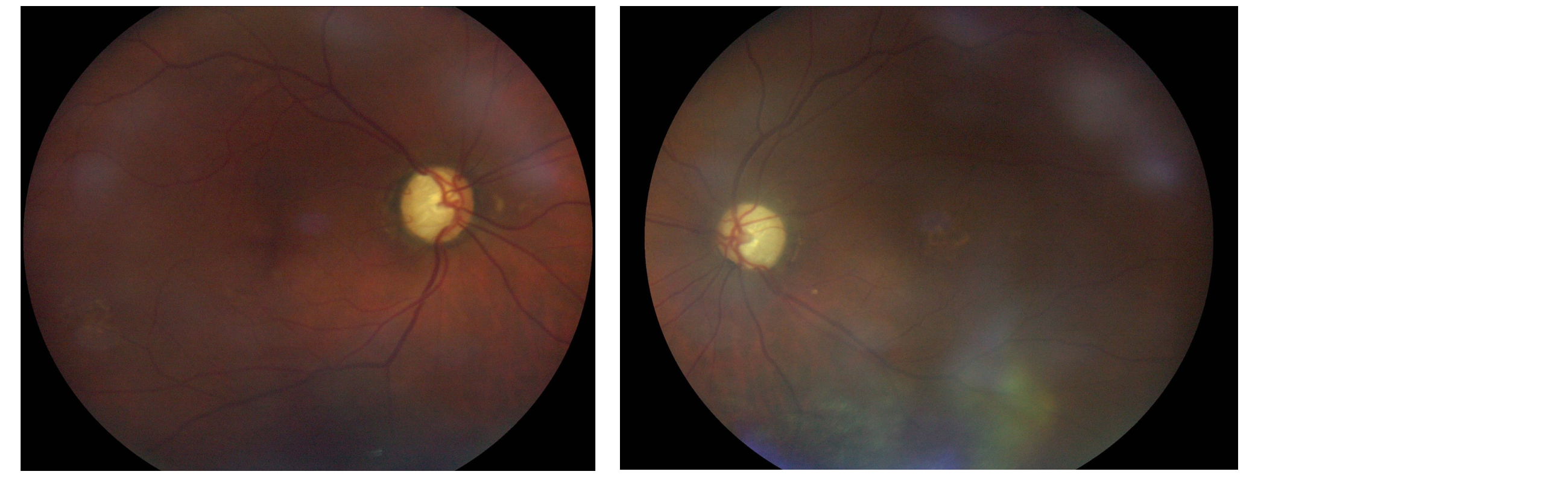
Figure 2:
Why Was an ERG Test Performed?
There was a prominent discrepancy between severe structural damage detected in OCT and fundus imaging as compared to surprisingly good central vision. Additionally, there was concern about minimal worsening in the cup-to-disc ratio from the last visit. In order to verify progression of the advanced glaucoma, ERG testing with the RETeval® was performed to provide an objective functional evaluation of retinal ganglion cell activity to compare to the structural findings.
What Were the ERG Findings?
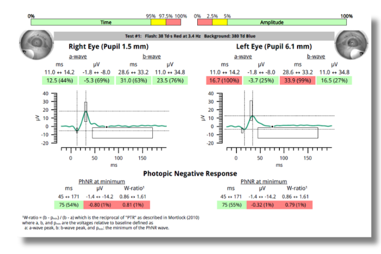
Initial RETeval testing (Figure 3) revealed the following:
| Right eye | Left eye | |
|---|---|---|
| b-wave Time | 32ms (89%) | 31.7 (85%) |
| W-ratio | 0.81 (1%) | 0.85 (2%) |
Figure 3: ERG at Baseline (September 2024)
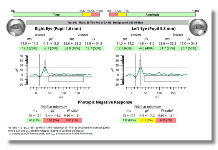
When the W-ratio is below 11% and/or b-wave time above 89% on the PhNR Report, the results are consistent with advanced glaucoma. In this case, the the W-ration was well below 11% in both eyes. At that time, the patient did not take topical glaucoma therapy, which was initiated at this visit. Three months after starting treatment, a follow-up ERG was performed (Figure 4), showing improvement in the b-wave time from 32ms (89%) to 31ms (63%) in the right eye. The W-ratio remained relatively stable at 0.81 in the right eye, however the left eye improved to 0.79. The W-ratio for both eyes remained in the abnormal range.
Figure 4: Follow-up ERG (January 2025)
How Did the ERG Impact Next Steps?
RETeval provided an objective, non-invasive way to monitor progression of ganglion cell function. In this patient, the ERG confirmed functional stability despite severe structural damage, even in the absence of visual field testing.
Specifically, the W-ratio remained stable from September 2024 to January 2025, with slight improvement in the W-ratio after initiating treatment. This helped to confirm effectiveness of the current treatment which was continued post ERG. Follow-up visits were scheduled every 4 to 6 months. ERG provided reassurance that retinal function was stable despite severe structural damage shown on OCT, validating our clinical decision-making.
Why We Use RETeval
In some glaucoma patients, visual field testing can be unreliable or the patient may refuse to take the test, as in this patient’s case. The RETeval handheld ERG, using the PhNR (Respuesta Fotópica Negativa) protocol, offers a valuable alternative to assess retinal ganglion cell function objectively. In this case, subsequent RETeval testing confirmed stable functional status and guided confident therapeutic decisions.
Josué Roberto Lozano, MD
Centro Médico Hidalgo (Monterrey, Nuevo León, Mexico)
Dr. Josué Roberto Lozano is a specialist in ophthalmology and subspecialist in retina and vitreous, based at Centro Médico Hidalgo in Monterrey, Nuevo León. A graduate of Universidad Autónoma de Nuevo León, he completed his specialization at Unidad Médica de Alta Especialidad #25 (tertiary level eye hospital with advanced medical services) and subspecialty training at private and public institutions. With over 10 years of experience, he also has advanced training in glaucoma and refractive cataract surgery. He is a member of the Mexican Retina Association, the Mexican Society of Ophthalmology, and the Ophthalmology College of Nuevo León.