Potencial Evocado Visual (PEV)
O que é o potencial evocado visual ( PEV)?
Como PEV funciona
Um estímulo luminoso fornecido por um monitor de computador ou uma tela de ganzfeld é recebido pelas células da retina do olho. O potencial elétrico viaja da retina até o córtex visual do cérebro. Eletrodos colocados no lóbulo occipital registram a atividade elétrica resultante no córtex visual.
O que ele nos diz
As anormalidades no PEV indicam possíveis distúrbios na via visual. O PEV pode ajudar a detectar o mecanismo causador dos déficits visuais.
- Ele é útil como um teste da via visual como um todo.
- Normal VEP indicates that visual information is successfully transmitted to the brain’s visual cortex for interpretation.
- As anormalidades podem indicar problemas com a transmissão do olho para o córtex visual.

O exame de PEV fornece dados funcionais e objetivos
O potencial evocado visual ( PEV) é uma medida não invasiva das respostas eletrofisiológicas do cérebro a estímulos visuais. O teste de PEV fornece informações funcionais objetivas e registráveis, mesmo para pacientes não cooperativos, não verbais ou inconscientes.
Embora a tecnologia de imagem, como a ressonância magnética, continue avançando, o PEV é considerado uma ferramenta valiosa para ajudar a detectar lesões ocultas na via visual e, principalmente, no nervo óptico.
Why conduct VEP testing?
O PEV é um teste objetivo da função visual. Diversas síndromes e anomalias podem afetar o PEV, inclusive neurite óptica, esclerose múltipla (EM), tumores, lesões traumáticas, infecções e agentes tóxicos. A interpretação das anormalidades do PEV pode ajudar a diferenciar os possíveis tipos de patologias subjacentes.
Aplicações clínicas do PEV
VEP, as an aid in diagnosis and disease management, is clinically valuable to clinicians in the determination of neurological deficits causing vision issues — even when imaging is unreliable or inconclusive, or when symptoms appear to be subjective only. Likewise, VEP can be used to track functional recovery after an acquired or traumatic neurological event.[44]
O teste de PEV pode, portanto, ser usado para avaliar:
- Desvio nas fibras do nervo óptico (por exemplo, albinismo)[43]
- Disfunções tóxicas ou nutricionais do nervo óptico[43]
- Suspeitas de neurite óptica (resultante de desmielinização, por exemplo, esclerose múltipla)[42]
- Recuperação de uma série de disfunções da via óptica[42]
- Cegueira cortical devido a meningite ou anóxia[42]
O teste de PEV também pode ser usado para:
- Distinguir entre inflamação e outras neuropatias ópticas[42]
- Determinar quantitativamente a função do sistema visual e as vias ópticas devido a traumatismo craniano[42]
- Ajudar a detectar tumores orbitais que comprimem a via óptica[42]
- Monitorar possíveis gliomas do nervo óptico em pacientes com neurofibromatose[42]
- Monitorar a função cerebral de pacientes em salas de cirurgia ou em terapia intensiva[44]
3 tipos de testes de PEV
Diferentes estímulos visuais foram projetados para produzir diferentes tipos de resultados de PEV. Em geral, os tipos de testes de PEV mais usados clinicamente são o PEV com reversão de padrão (PVEP ou PRVEP), o PEV com início de padrão e o PEV com flash. A estimulação reversa de padrão é o padrão ouro para o teste de PEV, com o tempo e a forma de onda mais consistentes.[45] On the other hand, pattern-onset VEP and flash VEP are more commonly used for patients who cannot fixate, such as infants.

PEV DE INVERSÃO DE PADRÃO
A forma de onda do PEV com reversão de padrão (PRVEP) é gerada por um painel de xadrez com reversão de fase. A forma de onda do PRVEP contém três picos de interesse: N70, P100 e N155. O P100 é a medida mais robusta, apresentando a menor variabilidade entre pacientes e entre olhos, além de alta reprodutibilidade.[47] Portanto, a robustez e a consistência do PRVEP permitem regras gerais de interpretação e são mais comumente usadas para pacientes com suspeita de doenças do nervo óptico.[43]
As indicações mais comuns em que o PEV PR é usado são neuropatia óptica traumática, neurite óptica, neuropatias ópticas isquêmicas ou disfunção tóxica ou nutricional do nervo óptico.[43][46] Essas condições são normalmente caracterizadas por formas de onda do PRVEP ausentes, relação de amplitudes reduzidas (olho afetado/olho amarelo) ou tempos de pico atrasados, com cada uma das indicações ajudando a identificar ou confirmar uma doença suspeita.[43]
However, VEP abnormalities are nonspecific, and an adjunctive test of macular function is required. This may include a pattern ERG or multifocal ERG[31] — conducted with the UTAS ou ERG flash[43] com o RETeval®.
PADRÃO-ONSET PEV
The pattern-onset VEP waveform is generated by an on-off checkerboard stimulation. The pattern-onset VEP is composed of C1-positive—C2-negative—C3-positive peaks. A core application of pattern-onset VEP is the detection of chiasmal misrouting of albinism in older patients, where this stimulus type is actually required.[45]
Também tem outras aplicações. Semelhante ao PEV Flash, o PEV Pattern-Onset é útil quando há nistagmo ou quando o paciente tem dificuldade de fixação (por exemplo, crianças pequenas). Os padrões de PEV estabelecidos pelo ISCEV reconhecem que, embora a variabilidade intrapaciente no PEV com flash e com padrão de início possa ser alta, as comparações interoculares dessas formas de onda podem fornecer informações clinicamente valiosas. Isso inclui a distribuição trans-occipital de respostas monoculares.
O versátil UTAS device can perform pattern-onset testing. However, for most applications, flash VEP can perform analogous testing functionality to that of pattern-onset. In this case, the handheld and portable RETeval pode ser uma solução melhor.

FLASH PEV
O PEV com flash é produzido por um flash branco gerado por uma taça ganzfeld. A forma de onda representa uma sequência de leituras negativas e positivas alternadas. Os picos P2 (correspondente ao P100 no PRVEP) e N2 são as medidas clinicamente mais relevantes.[44]
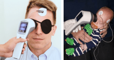
A principal vantagem do flash PEV é que não requer a cooperação do paciente para fixação/foco e pode ser usado mesmo quando o paciente está sedado. O flash PEV é, portanto, geralmente indicado em crianças pequenas, pessoas com deficiências de desenvolvimento ou pacientes que não cooperam com o paciente.[44]
O RETeval pode ser usado para monitorar a função cerebral mesmo em pacientes anestesiados ou em coma. A estimulação de Half-field pode indicar se as anomalias do PEV resultam de uma lesão retroquiasmática e, por esse motivo, é útil para ajudar no diagnóstico dos efeitos do albinismo.[44]
O dispositivo RETeval device is capable of performing flash VEPs.
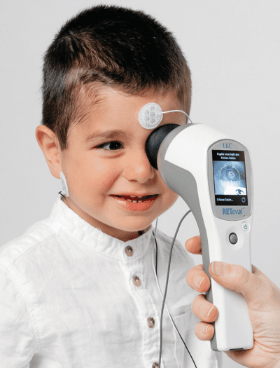
RETeval, testes PEV simplificados
More about the RETeval ERG/VEP device
Popular Topics: Make a Difference in Diabetic Retinopathy Care | Glaucoma Evaluation with RETeval PhNR Test | RETeval Device Reference Data | RETeval in Optometry
Case Studies: Photopic Negative Response as a Reliable Method for Glaucoma Follow-up in Children | A Tale of Two Patients | Vision Complaints Reflected on ERG | Predictive Value of Combining Diagnostic Technologies| ERG Provides Clarity When Fields and OCT Are Inconclusive| ERG Raises Red Flag, Changing Management Trajectory | ERG Provides Confidence to Monitor or Treat | ERG to Determine Ischemic Status | ERG to Replace FA for CRVO Treatment Decision | Using ERG to Monitor Glaucoma | Routine ERG Use Supports Complex Patient Management | ERG Alters Follow-up Schedule and Education for Patient with Diabetes | Using ERG for Management of Birdshot Chorioretinopathy | Using ERG to Monitor Glaucoma | Comprehensive Pediatric Assessment Using ERG in Challenging Cases | ERG Above and Beyond Retinal Imaging | ERG’s Role in Diabetic Retinopathy Progression Monitoring | Avaliação de Risco para CRVO baseado em ERG | ERG Supports Diagnostic Accuracy in a Pediatric Patient
Ebooks: Core Cases from the Clinical Compendium | Modern Fundamentals of Diabetic Retinopathy Management in Optometry | Elevating Patient Care with ERG
Articles: Electroretinography Added to AAO’s Diabetic Retinopathy Preferred Practice Pattern Guidelines | How Comfortable is the RETeval for Patients? |The Use of RETeval ERG/VEP in Pediatric Ophthalmology | How RETeval ERG Has Enhanced My Practice | The Use of RETeval ERG/VEP in Pediatric Ophthalmology | The Ultimate Guide to Diabetic Retinopathy in Primary Eyecare | What Type of Functional Testing Do You Prefer for Patients with Diabetes? | Major Milestone: RETeval Referenced in over 200 Publications | Collaboration to Elevate the Standard of Care for DR | Is ERG Needed if You Have Access to a Good Structural Imaging Device? | A Straightforward Approach to Managing and Supporting Patients with Diabetes | Simplify Grading and Risk Assessment in Diabetic Retinopathy | Simplify Daily Decision-Making with Modern ERG | Objective Functional Testing Needs in Diabetes and Glaucoma | Why Modern ERG is Re-Defining Diabetes Management | Diabetic Retinopathy Management Protocols for Optometry
Videos: RETeval: Function to Rely On | RETeval Handheld ERG: Features & Benefits | RETeval: Enhancing Collaborative Care | Handheld ERG for Primary Eyecare | Advice for Optometric Colleagues about Handheld ERG | Making a Difference in Diabetic Retinopathy Care | Changing the Way We Think About Electrodiagnostics | ERG Testing Made Simple | VEP Testing Made Simple | ERG Waveform | Introduction to Visual Electrophysiology | A Superior DR Progression Risk Assessment with the RETeval dispositivo | Improve Glaucoma Management with the RETeval Handheld ERG Device | Reshaping the Retinal Diagnostic Landscape | New Solutions for Infants with ROP | Using the RETeval in Myopia Research
Webinars: ABCs of ERG | ERG in Action | Blueprint for Functional Assessments | Predicting Vision Loss in a Busy Retina Practice | Objective, Functional Testing for Glaucoma? | Best Management Practices for Diabetic Retinopathy | How the RETeval Device Became a Daily Instrument in my Diagnostic Toolkit