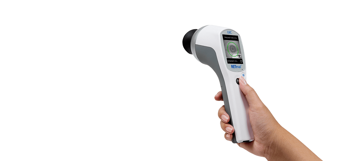
RETEVAL DEVICE FOR ELECTRORETINOGRAPHY
Revolutionary ERG/VEP
Anywhere for Anyone
Enhance your diagnostic capabilities or fuel your research with the RETeval® device — a powerful aid in the diagnosis and management of retina and optic nerve diseases such as diabetic retinopathy, glaucoma, and inherited retinal dystrophies.
Why RETeval?
For Clinicians

SIMPLE TO USE & INTERPRET
- Handheld, light, and portable
- Appropriate for any age without sedation
- Train technicians in minutes
- Mac and Windows Compatible
- Approved CPT code 92273 (USA)

ADVANCED TESTING
- Advanced full-field ganzfeld functionality in a handheld ERG/VEP device
- Simple to interpret
- Fully ISCEV-compliant
- 130+ peer-reviewed publications

NO CORNEAL CONTACT
- Our non-invasive patented Sensor Strip skin electrodes mean no corneal electrode required

DILATION NOT REQUIRED
- Real time pupillography adjusts for pupil size in real time – can be used in patients who are not dilated or in any stage of dilation
For Researchers

TRUSTED TESTS
- Access to the raw measurements as well as processed data
- Customize protocols with sinusoidal, square, ramp, and brief-flash stimuli
- RFF Extractor for exporting cursor values, electrical & pupil waveforms etc.

FUELING RESEARCH
- Non-invasive and highly-portable for maximum efficiency and compliance
- Achieve superior performance end-point tracking
- Ensure GLP and GCP with consistent, repeatable testing and results
VISUAL ELECTROPHYSIOLOGY TESTING
Detect Functional Stress
Anticipate Structural Damage
ERG and VEP tests provide objective information on the function of the visual system. It gives reliable guidance for medical professionals to manage functional changes that may impact a patient’s vision, typically in advance of structural changes.
Explore ERG Explore VEP
“We have been very pleased with the RETeval. It has changed the way that we practice and has allowed us to significantly decrease the amount of EUAs with ERGs we perform. Furthermore, it has resulted in earlier diagnosis of patients with inherited retinal degenerations.”

The RETeval device helps doctors to:
1. DETECT
Detect various retina and optic nerve diseases earlier
2. PREDICT
Predict the progression of the disease
3. FOLLOW
Then follow up on the course of disease
4. MONITOR
Monitor treatment success
Explore Applications
ERG and VEP play a vital role in obtaining diagnostic information in both humans and animals, aiding clinicians and researchers with comprehensive data on how the retina and visual pathways are functioning.

Diabetic Retinopathy
& Other Ischemic Conditions
The DR Assessment Protocol predicts the risk of progression. A score of 23.4 or higher means an 11-fold risk of requiring intervention within 3 years.

Glaucoma
& Other Optic Nerve Neuropathies
Aid and support the diagnosis and management of glaucoma by detecting and tracking changes to the ganglion cell layer.

Pediatrics
& Other Retinal Conditions
Identify dysfunction of rods and cones in children with suspected inherited retinal diseases with a non-invasive procedure, even in infants.
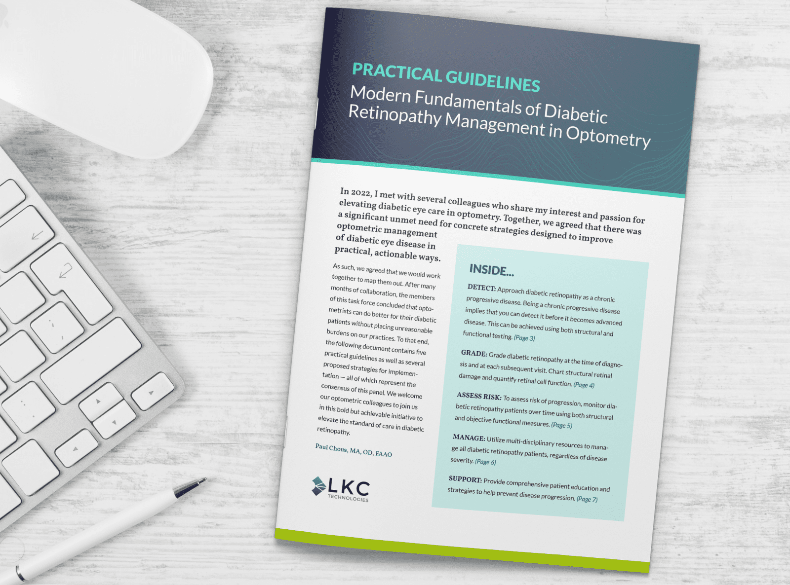
Modern Fundamentals of Diabetic Retinopathy Management in Optometry
A team of 14 optometrists set out to simplify and standardize diabetic retinopathy assessment and management by developing a set of practical guidelines for their peers. Their collaboration presents a framework to improve the diagnosis and management of DR to support improved patient outcomes and allow primary eye care providers to confidently care for the growing population of patients with diabetes.
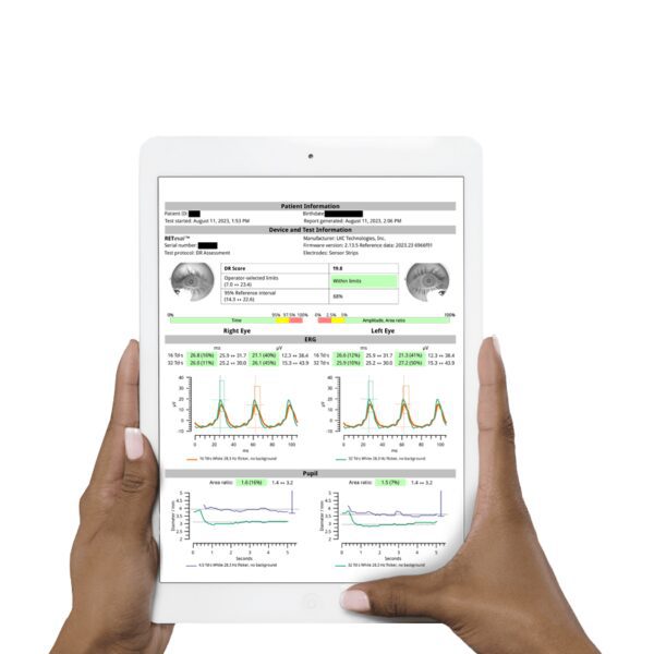
SIMPLE TO INTERPRET
RETeval Reports
Color-coded results compared to an age-adjusted reference data set make interpretation straightforward and efficient in a busy practice. Results are easily exported into any EMR/EHR system.
SAMPLE REPORTS
Diabetic Retinopathy Assessment
PhNR
ERG & VEP Coding and Reimbursement Guides
In the United States, there are more than 560 ICD-10-CM codes that may be associated with the CPT codes used to report electroretinography and visual-evoked potential. The following guides, developed by The Pinnacle Health Group, provide some of the more common diagnosis codes that may be used for protocols associated with the RETeval handheld device.
Download ERG guide Download VEP guide
The above guides focus on Physician Office Coding and Reimbursement. For Hospital Outpatient, contact us.
“RETeval really has made me a better diagnostician. It has allowed me to get early information about a patient’s eye health before disease becomes clinically visible.”

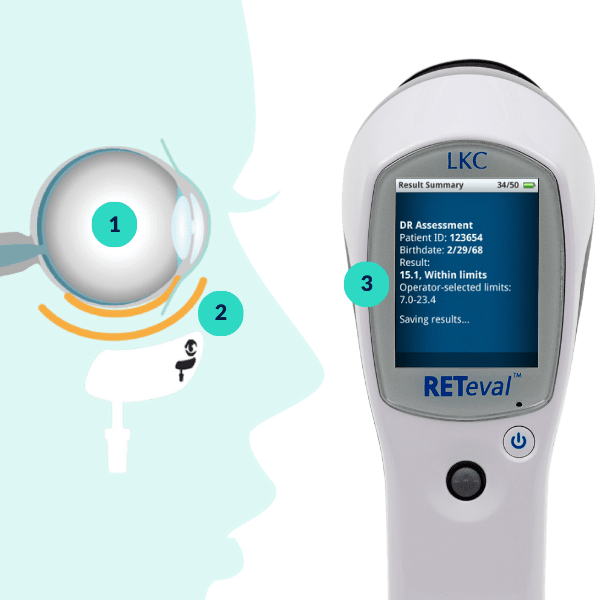
How It Works
-
The RETeval device starts flashing light into the patient’s eye.
-
The retina responds to the flashes by generating small electrical signals that travel through the facial structure to the Sensor Strip.
-
The Sensor Strip detects the electrical signals and compares the results to the age-adjusted reference database.
RETeval, ERG Testing Made Simple
RETeval Resources
RETeval Brochure (US)
RETeval Brochure (International)
Data Barcode App
ERG Coding & Reimbursement Guide (US)
CA Prop 65 Warning
Order Sensor Strips
Enhancing Risk Assessment in Patients with Diabetic Retinopathy by Combining Measures of Retinal Function and Structure
August, 2020
Brigell M, Chiang B, Maa A, Davis Q. Translational Vision Science & Technology. 2020; 9(9):40.
Screening for diabetic retinopathy in diabetic patients with a mydriasis-free, full-field flicker electroretinogram recording device
November 12, 2019
Zeng Y, Cao D, Yu H, et al. British Journal of Ophthalmology 2019;103:1747-1752.
Constant luminance (cd·s/m2) versus constant retinal illuminance (Td·s) stimulation in flicker ERGs
February 3, 2017
Davis Q, Kraszewska O, Manning C. Documenta Ophthalmologica. 2017: 134, 75–87.